
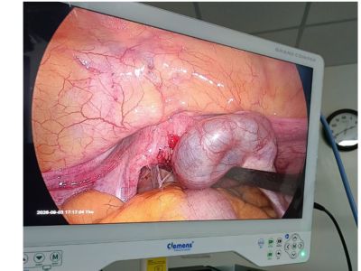
The patient presented with a history of primary infertility for 8 years. She had previously undergone evaluation for infertility, including hysterosalpingography (HSG), which showed bilateral tubal block.
The patient was also diagnosed with hyperthyroidism, which was first brought under control before proceeding with further fertility evaluation.
Considering the prolonged duration of infertility and bilateral tubal blockage, further evaluation with diagnostic hysteroscopy and laparoscopy was planned to identify the underlying cause and assess the pelvic anatomy.
The patient had been trying to conceive for approximately 8 years.
• The patient had been evaluated for primary infertility for approximately 8 years.
• HSG was performed, which showed bilateral tubal block.
• The patient was also diagnosed with hyperthyroidism, which was controlled before • proceeding with further fertility evaluation.
• No definitive treatment for the underlying pelvic adhesive disease had been performed before the current evaluation.
Suspected abdominal tuberculosis with genital tract involvement, associated with:
• Severe bilateral tubal disease
• Bilateral hydrosalpinx
• Dense pelvic adhesions
• Bilateral tubal blockage
• Extensive adhesions involving the uterus, intestine, pouch of Douglas and undersurface of the liver
• Hysterosalpingography (HSG): Bilateral tubal block
• Diagnostic hysteroscopy: Normal uterine cavity and bilateral tubal ostia; congested endometrium
• Diagnostic laparoscopy: Multiple pelvic and abdominal adhesions, bilateral hydrosalpinx, dilated and blocked tubes with clubbed ampullary ends
• Endometrial biopsy: Sent for TB NAAT and histopathological examination
• Diagnostic hysteroscopy
• Diagnostic laparoscopy
• Endometrial biopsy
• Bilateral tubal clipping
Bilateral tubal clipping was performed because of severe bilateral tubal disease and hydrosalpinx. The procedure aimed to exclude the diseased tubes from the uterine cavity and preserve the patient's future option of IVF after appropriate anti-tubercular treatment.
Duration: Evaluation and surgical management: Completed during the planned diagnostic and operative procedure. Further treatment: Appropriate anti-tubercular treatment is planned/considered based on biopsy and TB investigation results, followed by fertility reassessment.
• Diagnostic hysteroscopy
• Diagnostic laparoscopy for detailed assessment of pelvic anatomy
• Endometrial biopsy with TB NAAT and histopathological evaluation
• Bilateral tubal clipping as fertility-preserving surgical management before planned IVF
• Underlying pelvic pathology was identified that could not be fully explained by HSG alone.
• Severe bilateral tubal disease and hydrosalpinx were surgically addressed.
• The diseased tubes were excluded from the uterine cavity.
• The patient's future option of IVF was preserved following appropriate treatment and fertility planning.
Recovery Duration: Immediate post-operative recovery: Routine recovery following hysteroscopy and laparoscopy. Further fertility treatment: To be planned after completion of indicated treatment and reassessment.
The laparoscopic evaluation successfully revealed the likely underlying cause of the patient's long-standing infertility. Bilateral tubal clipping was performed because of severe tubal disease and hydrosalpinx. The patient now has a planned pathway toward IVF after appropriate anti-tubercular treatment and further fertility evaluation, preserving the possibility of achieving pregnancy in the future.
his case highlights how diagnostic laparoscopy can reveal the underlying cause of long-standing infertility when HSG alone cannot provide the complete picture.
Although HSG had demonstrated bilateral tubal block, laparoscopy revealed a much broader pattern of disease involving the pelvis, fallopian tubes and even the undersurface of the liver.
The combination of:
Extensive adhesions
Bilateral hydrosalpinx
Dense tubal adhesions
Clubbed ampullary ends
Pouch of Douglas involvement
Liver-surface adhesions
raised a strong clinical suspicion of abdominal tuberculosis with genital tract involvement.
This allowed further diagnostic testing through endometrial biopsy and helped guide fertility-preserving management.
Yes
The case involved:
8 years of primary infertility
Bilateral tubal block on HSG
Previous hyperthyroidism requiring control before fertility work-up
Diagnostic hysteroscopy and laparoscopy
Extensive pelvic and abdominal adhesions
Bilateral hydrosalpinx
Severe bilateral tubal disease
Suspected abdominal tuberculosis with genital tract involvement
Endometrial biopsy for TB NAAT and histopathology
Bilateral tubal clipping
Planned future IVF following appropriate anti-tubercular treatment
The most significant benefit was identifying the underlying pelvic pathology through laparoscopy and appropriately managing the severely diseased tubes while preserving the patient's future option of IVF.
This case demonstrates the importance of comprehensive evaluation in women with long-standing primary infertility and bilateral tubal block.
While HSG identified bilateral tubal obstruction, diagnostic laparoscopy revealed the extent and pattern of pelvic disease, including multiple adhesions, bilateral hydrosalpinx, dense tubal adhesions and adhesions over the undersurface of the liver. These findings were highly suggestive of abdominal tuberculosis with genital tract involvement.
An endometrial biopsy was therefore obtained for TB NAAT and histopathological evaluation.
In view of severe bilateral tubal disease and hydrosalpinx, bilateral tubal clipping was performed to exclude the diseased tubes from the uterine cavity and preserve the possibility of future IVF.
This case is a reminder that in women with prolonged infertility and bilateral tubal block, the diagnosis may not be limited to the tubes themselves—it may be revealed by the overall pattern of adhesions and pelvic anatomy seen during laparoscopy.