
• Pregnancy progressing normally until 32 weeks
• Baby's growth slowed on routine ultrasound
• Gradual reduction in amniotic fluid (Oligohydramnios)
• Progressive abnormal Doppler findings indicating reduced blood flow
• Increased risk of fetal distress due to placental insufficiency
• High-risk pregnancy requiring close monitoring
The pregnancy remained normal up to 32 weeks of gestation. During a routine ultrasound at 32 weeks, fetal growth restriction and decreasing amniotic fluid were detected. The condition was closely monitored until 37 weeks.
• Regular antenatal check-ups
• Routine ultrasound examinations
• Weekly ultrasound and Doppler monitoring after 32 weeks
• Continuous fetal growth assessment
• Close monitoring of amniotic fluid levels
• Careful observation for signs of fetal distress
• Fetal Growth Restriction (FGR/IUGR)
• Oligohydramnios (Low Amniotic Fluid)
• Progressively Abnormal Doppler Blood Flow
• Fetal Distress at 37 Weeks
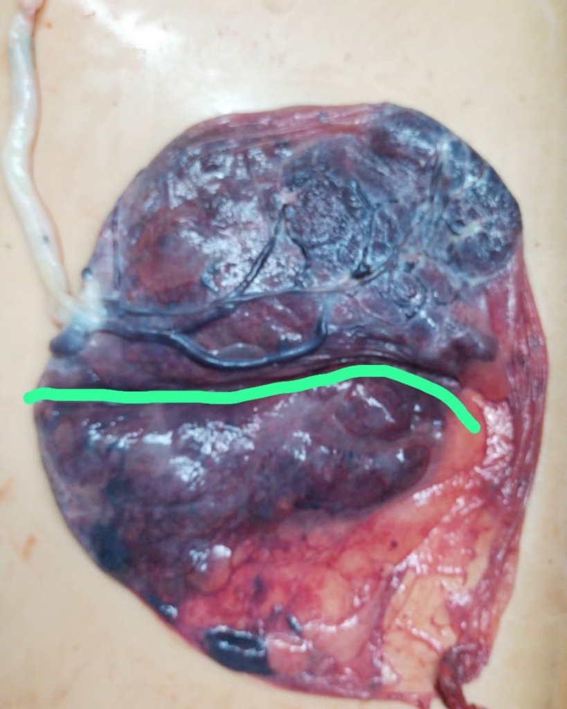
• Accessory Placental Lobe (Placenta Succenturiata) Detected During Cesarean Section
• Routine obstetric ultrasounds
• Serial fetal growth scans
• Weekly Doppler ultrasound studies
• Amniotic Fluid Index (AFI) assessment
Fetal heart rate monitoring
Intraoperative examination of the placenta
Emergency Lower Segment Cesarean Section (LSCS)
Duration: The patient was monitored closely from 32 weeks to 37 weeks of pregnancy. Emergency Cesarean delivery was performed Immediately after signs of fetal distress were detected.
• Serial fetal growth monitoring
• Weekly Doppler ultrasound surveillance
• Regular amniotic fluid assessment
• Early detection of fetal compromise
• Timely decision for emergency Cesarean delivery
• Detailed placental examination after delivery
• Healthy baby delivered safely
• Fetal distress managed before serious complications occurred
• Mother recovered well after Cesarean delivery
• No major maternal complications
• Baby received timely neonatal care
• Successful outcome despite a high-risk pregnancy
Recovery Duration: Mother recovered well during the routine post-operative period and was discharged in stable condition.
Early recognition of fetal growth restriction, falling amniotic fluid, and worsening Doppler findings enabled timely delivery, resulting in a safe outcome for both mother and baby.
Returned to Normal Activities: Yes
This was a rare obstetric case where the pregnancy appeared normal until 32 weeks. Careful weekly monitoring detected gradually worsening fetal growth, decreasing amniotic fluid, and abnormal Doppler changes. during emergency cesarean section placenta with an accessory placental lobe was discovered which was supplying to the baby.
• Fetal Growth Restriction (FGR)
• Progressive oligohydramnios
• Worsening Doppler blood flow abnormalities
• Fetal distress at term
Rare accessory placental lobe, known as placenta succenturiata
Timely emergency Cesarean delivery preventing serious fetal complications
Meticulous fetal surveillance using serial ultrasounds and Doppler studies enabled early detection of placental insufficiency and timely Cesarean delivery, ensuring the safety of both mother and baby.
This case highlights the importance of regular fetal growth scans, Doppler ultrasound monitoring, and timely clinical decision-making in high-risk pregnancies. Although the pregnancy progressed normally until 32 weeks, gradual fetal growth restriction, decreasing amniotic fluid, and abnormal Doppler findings signaled placental insufficiency. Emergency Cesarean delivery at 37 weeks revealed a rare accessory placental lobe, known as placenta succenturiata, which was supplying blood to the baby. Thanks to vigilant monitoring and prompt intervention by Dr. Hema Jajoo, both mother and baby had a successful outcome.